Want to read offline? [Download PDF]
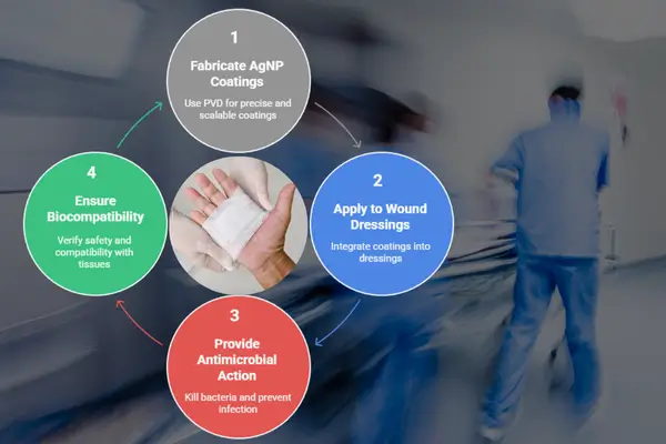
In the evolving field of advanced wound care, silver nanoparticle (AgNP) coatings have become a cornerstone for developing antimicrobial dressings that are both effective and biocompatible. Among various fabrication techniques, Physical Vapor Deposition (PVD) stands out for its ability to deliver precise, reproducible, and scalable silver coatings at the nanoscale.
This blog explores how PVD techniques are revolutionizing the application of silver nanoparticles in wound dressing materials, with a focus on deposition control, surface engineering, and antimicrobial efficacy.
Why Silver Nanoparticles?
Silver has long been known for its broad-spectrum antimicrobial properties. When reduced to the nanoscale, silver’s surface area increases dramatically, enhancing its ability to:
- Disrupt bacterial membranes
- Generate reactive oxygen species (ROS)
- Bind bacterial DNA
- Inhibit biofilm formation
This makes AgNPs ideal candidates for wound dressings, especially in treating chronic wounds, burns, and diabetic ulcers, where infection control is critical.[1]
The Role of PVD in Nanoparticle Coating
Physical Vapor Deposition (PVD) is a vacuum-based coating method where materials are vaporized and condensed onto a substrate. In the context of wound dressing applications, PVD offers precise control over silver nanoparticle size, distribution, and coating thickness, all of which are vital for consistent antimicrobial performance.[2]
Key Advantages of PVD for Silver Nanoparticle Coatings:
- Ultra-clean deposition (no solvents or binders)
- Controllable particle size and loading
- Strong adhesion to polymer or textile substrates
- Uniform coverage over large areas
- Scalable for industrial production
PVD Techniques for Nanoparticle Synthesis
There are several PVD-based techniques tailored for nanoparticle deposition:
- Gas-Phase Aggregation Sources : Specialized sources like Nikalyte’s NL-UHV nanoparticle source use gas-phase condensation to generate nanoparticles from ultra-pure solid silver source material, which are then deposited onto a wound dressing substrate.[3]Coatings deposited using gas phase aggregation incur a very low heat load onto the surface. This allows for:
- Controlled nanoparticle size (typically 2–20 nm)
- Narrow size distribution
- Adjustable surface coverage (mg/cm² scale)
- Coating of heat sensitive substrates.
- Magnetron Sputtering : Silver nanocoating can also be generated using Magnetron sputtering. When the coating thickness is less than 1 monolayer, aggregation of the silver atoms on the surface forms a rough high-purity nanotextured coating. However, control over the nanofeature size and loading is poor.[4] Magnetron sputtering also has the added disadvantage that the heat load on the substrate can be high due to direct exposure to the plasma, so heat sensitive substrate may not be compatible with this process.
- Thermal Evaporation : can also be used to generate silver thin film coatings with antimicrobial properties.[5]
Surface Engineering for Wound Dressings
Using PVD, silver nanoparticles can be deposited on various biocompatible substrates such as:
- Nonwoven polymer meshes (e.g., PU, PET, PCL)[6]
- Hydrogels[7]
- Electrospun nanofibers[8]
- Textile bandages[9]
The deposition process can be tuned to achieve monolayer or multilayer coatings, or gradient structures where silver content varies with depth helping to balance antimicrobial efficacy with cytocompatibility.[10]
Antimicrobial Efficacy & Biocompatibility
Studies have shown that PVD-deposited AgNPs:
- Inhibit a wide range of pathogens (e.g., S. aureus, E. coli, P. aeruginosa)
- Maintain long-term antimicrobial activity due to slow, controlled Ag⁺ ion release
- Minimize cytotoxicity by avoiding excess silver loading
- Remain stable and active under physiological conditions
Importantly, the absence of chemical stabilizers or surfactants in PVD-deposited nanoparticles makes them especially suited for medical applications.[11]
Scalability & Regulatory Considerations
PVD systems can be scaled from lab-scale prototype coatings to pilot and industrial production, ensuring the feasibility of commercial silver nanoparticle-coated wound dressings. Regulatory agencies such as the FDA and EMA require detailed material characterization, which PVD supports by producing coatings with consistent composition, thickness, and release profiles, key for regulatory approval.[12]
Nikalyte’s PVD Solutions for Silver Nanoparticle Coatings
At Nikalyte, we offer a range of PVD systems and instruments specifically designed for silver nanoparticle deposition, ideal for wound dressing applications. Whether you’re conducting early-stage research or scaling up for production, our solutions are built to support every stage of development. Our benchtop nanoparticle recipe-driven systems are perfect for academic labs and R&D teams simple to use, fully automated, and optimized for repeatable results, making them ideal for use across cross-functional research teams. For industrial needs, we provide scalable, high-throughput systems capable of uniform coating across large surface areas, making mass production of antimicrobial wound dressings both practical and efficient.[13]
Conclusion
PVD-based silver nanoparticle coatings offer a clean, scalable, and controllable method for enhancing wound dressings with potent antimicrobial properties. As wound care moves toward more advanced, multifunctional materials, PVD provides a robust platform for integrating silver nanotechnology into next-generation medical devices.
Contact us to speak with a technical expert about depositing silver nanoparticle coatings for wound care applications using our advanced deposition systems and instruments.
Reference
- Meenalotchani, R., Nirenjen, S., Manisha, M., Prajapati, B. G., Arun, E., Afreen, N., Ankul, S. S., Sridevi, S., Harikrishnan, N., & Alsaidan, O. A. (2025). Recent advances in silver nanoparticles for enhanced wound healing with mechanisms, formulations, and clinical insights. Journal of Drug Delivery Science and Technology, 83, 107437. https://doi.org/10.1016/j.jddst.2025.107437
- Ramachandran, K., Gaidi, M., Columbus, S., Daoudi, K., & Hammouche, J. (2023). Fabrication of noble metal–based antimicrobial nanosystems. In Antimicrobial Nanosystems: Fabrication and Development (pp. 353–375). Micro and Nano Technologies. https://doi.org/10.1016/B978-0-323-91156-6.00023-3
- Huttel, Y., Martínez, L., Mayoral, Á., & Fernandez-Martinez, I. (2018). Gas-phase synthesis of nanoparticles: Present status and perspectives. MRS Communications, 8(3), 1–8. https://doi.org/10.1557/mrc.2018.169
- Musa, I., Qamhieh, N., & Mahmoud, S. T. (2023). Ag nanocluster production through DC magnetron sputtering and inert gas condensation: A study of structural, Kelvin probe force microscopy, and optical properties. Nanomaterials, 13(20), 2758. https://doi.org/10.3390/nano13202758
- Le Thi Tam, L., Phan, V. N., Lan, H. T. T., Thuy, N. T., et al. (2013). Characterization and antimicrobial activity of silver nanoparticles prepared by a thermal decomposition technique. Applied Physics A, 113(3). https://doi.org/10.1007/s00339-013-7810-4
- Santo, D., Castro, J. D., Cruz, S., Carvalho, I., Cavaleiro, A., & Carvalho, S. (2025). Customisation of PVD coatings for biomedical devices. Surface and Coatings Technology, 512, 132277. https://doi.org/10.1016/j.surfcoat.2025.132277
- Popescu, I., Constantin, M., Solcan, G., Ichim, D. L., Rata, D. M., Horodincu, L., & Solcan, C. (2023). Composite hydrogels with embedded silver nanoparticles and ibuprofen as wound dressing. Gels, 9(8), 654. https://doi.org/10.3390/gels9080654
- Pagnotta, G., Graziani, G., Baldini, N., Maso, A., Focarete, M. L., Berni, M., Biscarini, F., Bianchi, M., & Gualandi, C. (2020). Nanodecoration of electrospun polymeric fibers with nanostructured silver coatings by ionized jet deposition for antibacterial tissues. Materials Science and Engineering: C, 113, 110998. https://doi.org/10.1016/j.msec.2020.110998
- Tahir, I., Amina, S. J., Ahmad, N. M., & Janjua, H. A. (2024). Antimicrobial coating of biologically synthesized silver nanoparticles on surgical fabric and surgical blade to prevent nosocomial infections. Heliyon, 10(17), e35968. https://doi.org/10.1016/j.heliyon.2024.e35968
- Krishnan, P. D., Banas, D., Durai, R. D., Kabanov, D., Hosnedlova, B., Kepinska, M., Fernandez, C., Ruttkay-Nedecky, B., Nguyen, H. V., Farid, A., Sochor, J., Narayanan, V. H. B., & Kizek, R. (2020). Silver nanomaterials for wound dressing applications. Pharmaceutics, 12(9), 821. https://doi.org/10.3390/pharmaceutics12090821
- Tabassum, N., Khan, F., Jeong, G.-J., Jo, D.-M., & Kim, Y.-M. (2024). Silver nanoparticles synthesized from Pseudomonas aeruginosa pyoverdine: Antibiofilm and antivirulence agents. Biofilm, 7, Article 100192. https://doi.org/10.1016/j.bioflm.2024.100192
- Saad, K. S. K., Saba, T., & Rashid, A. B. (2024). Application of PVD coatings in medical implantology for enhanced performance, biocompatibility, and quality of life. Heliyon, Article e35541. https://doi.org/10.1016/j.heliyon.2024.e35541
- Yudaev, P., Mezhuev, Y., & Chistyakov, E. (2022). Nanoparticle-containing wound dressing: Antimicrobial and healing effects. Gels, 8(6), 329. https://doi.org/10.3390/gels8060329